Contact Us or call 800-838-4268 for more information
Skip to main content

Feline Pancreatitis

Patient Information

Age
10 Years
Gender
Female, Spayed
Species
Feline

Images

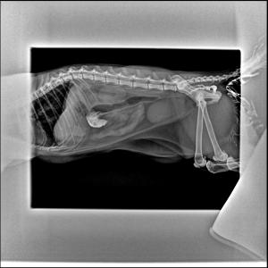
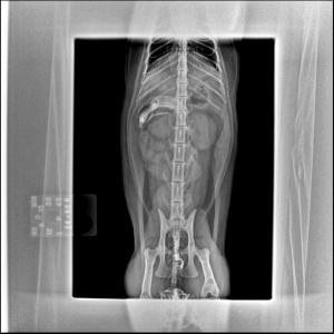
Minor retention of barium is noted in the transverse colon. Otherwise no significant abnormalities are present.
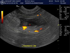
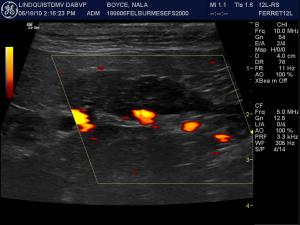
Power Doppler assessment of the pancreatic parenchyma demonstrates subnormal blood flow consistent with some level of avascular necrosis.
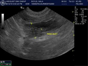
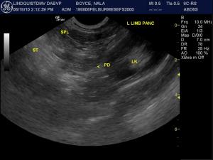
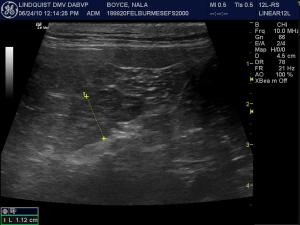
Still image of the left pancreatic limb demonstrating excessive width (1.48 cm, N.L. < 0.8 cm) and deviated pancreatic duct (central).
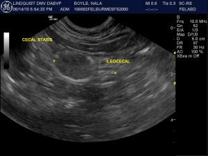
Ileo-cecal region demonstrating cecal stasis, a common finding in systemically sick patients with diminished Gi motility. This is not to be confused with an “obstructive pattern” given the distal ileum to the right and cecum to the left.
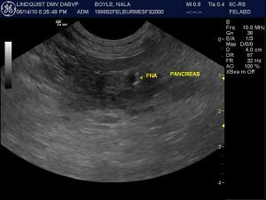
22-gauge fine needle aspiration precisely placed into the hypoechoic pancreatic pathology to maximize sampling accuracy. The hypoechoic non-vascular regions are most accurate representations of the pathology in act in my experience. Sampling multiple regions of varying sonographic echogenicities helps improve the “global” representation of the pathology for the cytologist when performing FNA or histopathology when performing tru-cut biopsies.
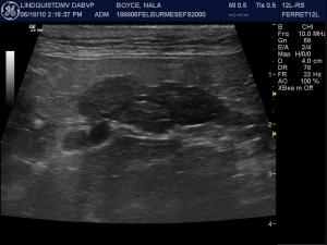
Follow-up sonogram after 4 days of intensive care reveals increased echogenicity compared to the dramatic hypoechoic images at initial presentation. The patient was less painful regarding the probe pressure in this region.
Same lesion as images 12/13 but with high resolution linear probe demonstrating duct (central) and capsular deviation consistent with chronic retraction owing to fibrosis, The patient was mildly painful upon imaging indicating persistent active inflammation and edema. However, the width of the pancreatic limb has diminished corresponding to the clinical improvement. The lesion is now more localized and sectorial compared to the diffuse left limb pathology noted at the initial presentation.
Power Doppler assessment of the follow-up presentation demonstrates subnormal blood flow to the residual hypoechoic region of the left pancreatic limb.
3-week follow-up of the same left pancreatic limb demonstrating residual excessive thickness and parenchymal remodeling. The patient was no longer painful in this region however. Clinical signs were essentially resolved at this point. Undulating capsule was now noted consistent with fibrous retraction and a common finding as sequelae to an inflammatory event.

History

Feline Pancreatitis (“Il Grande Mistero”), the sonogram, & the needle. Cutting to the chase in feline pancreatic pathology.

Case managed by Dr. Erno Hollo, Dr. Keith Ross & staff at Basking Ridge Animal Hospital, Basking Ridge, NJ, USA. Sonogram and diagnostic evaluation performed by Eric Lindquist DMV (Italy), DABVP (Canine & Feline Practice), NJ Mobile Associates & SonoPath.com.?

Sonogram: Nala

History: (Colleen Puelsch BS, Kelly Vasquez RVT)): An 10-year-old FS Burmese cat was presented for anorexia, vomiting, and soft stool. The physical exam revealed mild dehydration, tacky mucous membranes, fever of 104 degrees F, and mild cranial abdominal pain upon palpation. CBC and blood chemistry analysis revealed only mild elevation in triglycerides. fPLI and urinalysis were not performed.

Clinical Differential Diagnosis

(Remo Lobetti PhD, DECVIM): GI tract - IBD/neoplasia/foreign body Pancreas - pancreatitis/neoplasia/cyst/abscess Liver - cholangio-hepatitis/hepatitis/neoplasia Peritonitis

Image Interpretation

(Lindquist DMV, DABVP)

Sonographic Differential Diagnosis

(Lindquist DMV, DABVP): Pancreatic necrosis/pancreatitis with possibility of pancreatic carcinoma, lymphoma, or nodular hyperplasia.

Sampling

(Lindquist DMV, DABVP): US-guided fine needle aspiration of multiple areas of the pancreas revealed well differentiated pancreatic cells with increased neutrophils admix with blood consistent with suppurative inflammation. US-guided fine needle aspiration of the spleen also revealed suppurative inflammation.

Outcome

The patient was stable as an outpatient after 1 month of zithromax and hypoallergenic diet.

Comments

At the time of this on line presentation, the author is unaware of any studies evaluating cytology or U/S guided biopsies of the pancreas and they are not done routinely. However, the author has performed the procedure multiple times without any complications. It is imperative when performing FNA or biopsies that multiple regions of the organ be aspirated/biopsied due to the risk of missing a very localized lesion, which is very common with the pancreas. There are a number of ultrasonographers, including the author, who have performed the procedure multiple times without any complications (personal communications).

Videos

The left limb of the pancreas is coarse and excessively hypoechoic compared to surrounding omentum. Limb enlargement is present with a width greater than 1 cm and hypoechoic extention of the tissue through at least 3 cm of the left pancreatic limb. Dilation of the pancreatic duct is noted in its normal central location. Deviation from curvilinear capsular and duct contour is noted. The patient demonstrated focal pain upon imaging of this region but tolerated the probe pressure readily in other regions of the abdomen.
Video of another region of the left pancreatic limb demonstrates better power signal uptake in portions of the pancreatic tissue.
Video utilizing harmonics to help improve resolution of the pathology and eliminate unnecessary artifact.
Video of the ileo-cecal valve demonstrating the ileum being “driven” into the cecum to differentiate this from an obstructive pattern.
Video of FNA of the pancreas. Given that there is little room for sampling (1-1.5 cm width of pathology), short jabs are taken followed by a slight twist of the syringe (“corkscrew technique”) during the jab.
Minor uniform splenic enlargement was noted. Hence, US-guided FNA was also performed to define underlying cytopathology.
10-days follow-up of the same patient after 1 week of IV fluid therapy, zithromax, Gi protectants and nutritional support. Significantly diminished pathological volume is evident in the left pancreatic limb. The patient was only minimally painful upon imaging during this session. A 1 cm width x 2 cm length of residual pathology is evident.
Same lesion as images 12/13 but with high resolution linear probe demonstrating duct (central) and capsular deviation consistent with chronic retraction owing to fibrosis, The patient was mildly painful upon imaging indicating persistent active inflammation and edema. However, the width of the pancreatic limb has diminished corresponding to the clinical improvement. The lesion is now more localized and sectorial compared to the diffuse left limb pathology noted at the initial presentation.
Power Doppler assessment of the follow-up presentation demonstrates subnormal blood flow to the residual hypoechoic region of the left pancreatic limb.
3-week follow-up of the same left pancreatic limb demonstrating residual excessive thickness and parenchymal remodeling. The patient was no longer painful in this region however. Clinical signs were essentially resolved at this point. Undulating capsule was now noted consistent with fibrous retraction and a common finding as sequelae to an inflammatory event.