Contact Us or call 800-838-4268 for more information
Skip to main content

Gastro-Enteritis

Patient Information

Age
3 Years
Gender
Male, Neutered
Species
Canine

Images

History

Surprise, surprise, surprise! After 2 months of GI foreign bodies on ultrasound, take a look at this foreign body that was not in the stomach and this is no April fools trick.

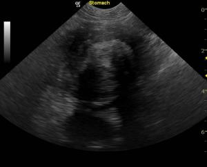
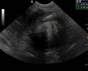
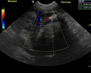
Sonogram (Pancreas): Gunner (Name changed to protect the innocent :)

History: A 3-year-old, NM Pointer cross was presented for vomiting/diarrhea. Endoscopy of the stomach and duodenum was within normal limits. Blood work was unremarkable and palpation of the cranial abdomen revealed some discomfort.

Clinical Differential Diagnosis (Lobetti):

GIT – non-specific gastro-enteritis (viral, bacterial, helminths, protozoa, dietary indiscretion, toxins), dietary hypersensitivity, IBD, foreign body, emerging lymphoma
Pancreas – pancreatitis, abscess
Addison's disease

Sonographic Interpretation (Lindquist):

The gastrointestinal tract presented relatively normal mucosa with an empty lumen.

Sonographic Differential Diagnosis

Sonographic Differential Diagnosis (Lindquist): Large pancreatic abscess with peri pancreatic inflammation and gastrointestinal impingement. Spasmic duodenum and duodenitis. There is the possibility for foreign body embedded within this abscess given the organization. At time foreign bodies can pass through the intestinal wall and embed into the abscess. Surgical intervention would be recommended; however, it will be difficult to resect the lesion completely. Therefore, removal of as much necrotic debris as possible and omentopexy would likely be the intervention of choice. Guarded prognosis.

Sampling

Surgical intervention: ex-lap.

Outcome

A gauze sponge was removed surgically without complication and debridement of necrotic tissue was performed. The patient had undergone gastrointestinal surgery in the past at another facility but the history was unclear. The patient recovered uneventfully.

Videos

The axis of the pyloric antrum was deviated from a large progressively shadowing pancreatic mass or abscess/necrosis occupying the right pancreatic base. It measured approximately 4.0 cm with shadowing material within the lumen. Peri-pancreatic inflammation and acute on chronic mixed echogenic tissue changes were also noted associated with the pancreas.
Hi resolution linear image demonstrates spasming of the proximal duodenum adjacent to the lesion with periserosal and peripancreatic ill-defined echogenic fat suggestive for inflammation.