Contact Us or call 800-838-4268 for more information
Skip to main content

Grass Awn in a 2-Year-Old MN Golden Retriever: Our Case Of the Month December 2019

Patient Information

Age
2 Years
Gender
Male, Neutered
Species
Canine

Keywords

Images

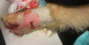
Grass Awn in front left foot.
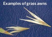
Examples of grass awns.
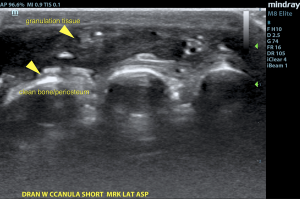
Clean bone/periosteum.
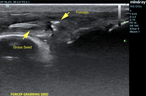
Forceps grabbing onto grass awn.
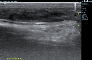
Image post removal of grass awn.
Front left foot.
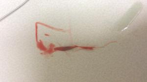
Grass awn removed from front left foot.
Grass Awn vs. Ultrasound … Who will win?
 
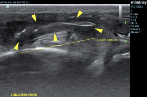
Spring & fall are great times of year, except for those pesky grass awns that like to get everywhere, and we mean EVERYWHERE!   Grass awn foreign bodies are very common, especially in the Pacific Northwest. This plant material can be inhaled or penetrate through the skin and migrate elsewhere, and backward-pointing barbs generally prevent effective elimination without intervention.  Swellings, draining tracts and abscesses commonly occur when the paws are affected, as is the case here.  Ultrasound is ideal to have in your toolbox for these “out of the box” presentations, when this or any foreign body is on the differential list.
 
Thank you to Dr. Eric Glaze of Albany Animal Hospital for asking us to join him in this grass awn seeking expedition with his sweet golden retriever patient.  This foreign body retrieval was made possible with the studious efforts of Amanda Lacey, SDEP™ Certified clinical sonographer of Animal Sounds Northwest
and SonoPath specialist, Dr. Eric Lindquist, DMV, DABVP, Cert. IVUSS for his "on-site" telemedicine interpretation via video chat. 

History

The patient was presented for drainage tract between the 2nd and 3rd digits in the front left foot. Previous probing for the grass awn was unsuccessful; an ultrasound was performed to aid in the process of pinpointing the location of the grass awn and image the surrounding tissues.

Image Interpretation

A front leg soft tissue lesion was imaged in this patient. Granulation bed was noted and a grass awn foreign body was evident, measuring approximately 1.5 cm. Regional thickened tissue was noted and minor fluid was noted within the cavity. The periosteum did not appear affected. Soft tissue cellulitis pattern was noted. Regional joints were unaffected.  Culture of the granulation tissue would be ideal. Recheck sonogram if wound healing is not adequate over the next 1-2 weeks.

DX

Grass awn granuloma/fistula

Outcome

An onsite ultrasound-guided surgical cut-down was performed and complete removal of the grass awn was successful. The patient has since recovered with no complications.

Videos

Grass awn.
Forceps grabbing grass awn.