Ultrasound a stifle? Sure … Why not? When image protocols are defined by Dr. Nele Ondreka DECVDI (http://www.sonopath.com/about/specialists/dr-nele-ondreka-dipecvdi) from the University of Giessen (Germany), one of the early founding institutions of orthopedic ultrasound, then partial and total cruciate ruptures, bucket handle meniscal tears, degenerative and proliferative inflammatory and degenerative articular and bone lesions are all part of the fun when you have a limb and a probe. It's all about the protocol and Dr. Tim Hunt of Bayshore Vet in Marquette, Michigan, America’s Favorite Veterinarian 2014, is one SonoPath client that took to this technique like one of his sled dogs to a bone :). Fresh off a 36th place finish at the Iditarod this year, Tim is on the knee with a probe in the Michigan UP. See what Tim and Nele found in this knee in October 2015 SonoPath case of the month! The dx efficiency to stifles: Scan it, diagnose it, cut it, confirm it, cure it:)
Image Interpretation
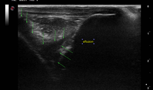
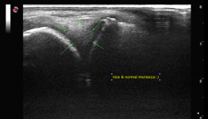
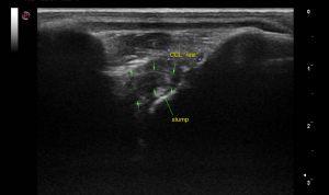
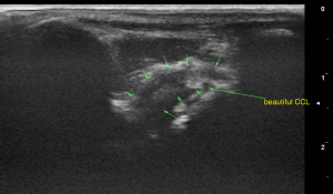
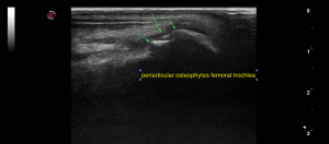
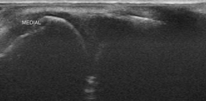
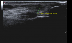
The left stifle joint presented moderate anechoic effusion, moderate capsular thickening and moderate synovial proliferation. There were mild to moderate osteophyte formations at the periarticular margins. The femoral condylar cartilage surface appeared to be mildly roughened. The cranial cruciate ligament (CCL) presented partial loss of integrity with irregular margination, uneven thickness and periligamentous effusion. There was a small hyperechoic fibre stump visible next to the distal insertion of the CCL at the tibial intercondylar eminence. The caudal cruciate ligament was not seen. The medial and lateral meniscus did not present ultrasonographic abnormalities.
Sampling
Surgery confirmed partial tear of the cranial cruciate ligament and the meniscus was in tact.
DX
The ultrasonographic findings are compatible with a left-sided partial CCL rupture and chronic degenerative joint disease with moderate osteoarthrosis and synovitis.

