Contact Us or call 800-838-4268 for more information
Skip to main content

Mural Intestinal Abscess In A 2-Year-Old MN Doberman Pinscher: Our Case Of the Month June 2022

Patient Information

Age
2 Years
Gender
Male, Neutered
Species
Canine

Images

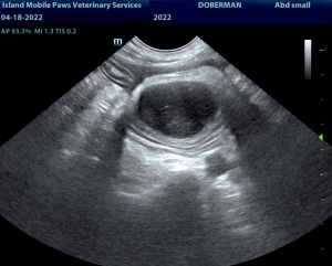
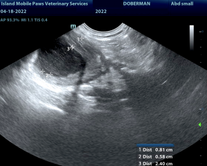
Mural intestinal abscess location cranial to the urinary bladder.
Mural intestinal abscess.
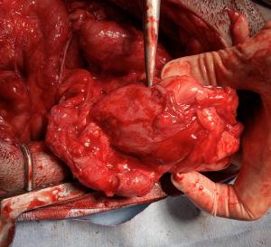
Mass found during exploratory surgery.
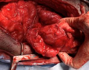
Jejunum entwined around mass.
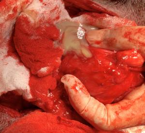
Mass opened revealed pus.

Doberman Pinscher + 1 sock = FB + exploratory x 1 month = same Doberman + yet another sock = 2nd FB + exploratory x adhesions = intestinal abscess. Dr. Donna Markland, owner of Island Mobile Paws Veterinary Services (serving the Cowichan Valley and Saltspring Island areas) provided the diagnostic imaging and work up for this mischievous Doberman with a taste for socks. Detailed interpreation by Eric Lindquist, DMV, DABVP, Cert. IVUSS owner of SonoPath.com.

History

A 2-year-old MN Doberman Pinscher presented on April 4th due to a sock foreign body. He underwent exploratory surgery with a resection and anastamosis. This was the second resection and anastomosis surgery for this patient since February. After the first surgery, the patient had a septic abdomen. After that surgery, he was managed with a drain in an emergency hospital for several days. He had initial hepatic enzyme elevations which resolved with resolution of sepsis. At the second surgery on April 4th, adhesions were noted throughout the abdomen. There was an area of adhesions in the left inguinal area that was extremely friable. A drain was placed following this surgery as well. Intracelluar bacteria were noted in the drainage fluid. The patient was discharged from the hospital on April 8th. The following medications were sent home: Cefaseptin, 750 mg BID x 5 days Baytril, 150 mg BID x 5 days, Metronidazole, 500 mg BID x 5 days Cerenia and codeine were also dispensed. Clinically the patient is doing very well. He is eating, drinking, urinating and defecating normally. His activity level is good. Blood work from April 7th: Alb=21 (better than pre-op) ALP=241, lymphocytes=0.88, monocytes=1.57, Neutrophils=12.15, suspected bands. A follow up ultrasound 14 days post-op from the second sock foreign body surgery was performed.

Image Interpretation

The upper gastrointestinal tract was unremarkable. A 3.0 cm abscess was noted deriving from what appears to be the intestinal wall with regional peritonitis. Some adhesions were noted with deviation of the normal intestinal contour. Localized free fluid was noted. This appears to be isolated and deriving from the jejunum.

Sampling

Sample of the pus from the mass was obtained during exploratory surgery and sent out for culture.

DX

Mural intestinal abscess with regional peritonitis.

Outcome

Immediate exploratory surgery was recommended with resection and abdominal lavage; neoplasia is unlikely. The patient was referred to an emergency hospital for surgery. Notes from the exploratory: There was not any free fluid in the abdomen. There was a massive amount of adhesions from mid-abdomen caudally. Many of the adhesions were broken down. A 5cm mass was located on the right mid abdominal wall. The jejunum was entwined around the mass. The mass was opened. About 15 to 12 mls of pus exuded from it.The pus was cultured. The interior of the mass was explored. There was not any foreign body found within it. The mass could not be removed due to the intestines entwining with it. The mass was lavaged and a section of the omentum was transferred to the cavity with the mass and sutured into place. The abdomen was lavaged liberally. Pathology results from the pus found Escherichia coli - 4+, Klebsiella variicola - 3+, Enterococcus faecalis - 4+.

Videos

Mural intestinal abscess.
Doppler of abscess.