Contact Us or call 800-838-4268 for more information
Skip to main content

Obstructive urinary disease part 1: In a 12-year-old MN Beagle. Our Case Study Of The Month June 2014

Patient Information

Age
12 Years
Gender
Male, Neutered
Species
Canine

Images

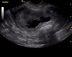
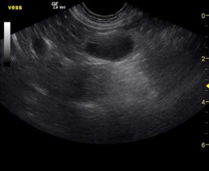
Image 1: The urinary bladder presented a concentric, parenchymal mass that occupied most of the apical ventral and apical dorsal aspects.
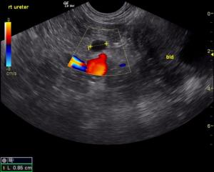
Image 2: Right hydroureter.
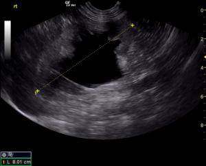
Image 3: Right kidney with moderate to severe hydronephrosis.
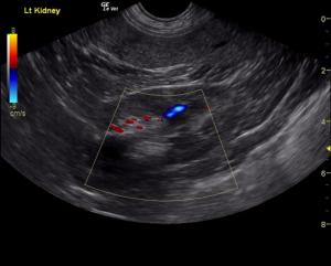
Image 4: The left kidney measured demonstrates minor degenerative changes.
Image 5: The iliac lymph node was enlarged

Why do our patients feel like a truck driver waiting to cross Governor Christie's George Washington bridge? https://www.youtube.com/watch?v=VKHV0LLvhXM

History

A 12-year-old MN Beagle weighing 50 lbs, was presented for hematuria after no improvement on medications. Urine specific gravity was 1.010, pH 7.1, protein 3+, blood 4+.

Clinical Differential Diagnosis

Urinary/prostatic neoplasia, urolithiasis, UTI, coagulopathy, idiopathic.

Image Interpretation

The urinary bladder presented a concentric, parenchymal mass that occupied most of the apical ventral and apical dorsal bladder with extensive polypoid changes entering into the caudal aspect of the ventral bladder and cystourethral junction. Dorsal polyps were also noted. Right hydroureter was noted which extended proximally to the right kidney with moderate to severe hydronephrosis. Hydronephrosis measured 3.82 x 4.0 cm. The right kidney measured 8.01 cm in length. The left kidney measured 7.0 cm with minor degenerative changes. No obstructive disease was noted. The iliac lymph node was enlarged in this patient, hypoechoic and irregular measuring approximately 2.0 cm. This is strongly suggestive for metastatic disease. Other small iliac lymph nodes were enlarged.

Sonographic Differential Diagnosis

Concentric bladder mass occupying most of the bladder with right, obstructive hydroureter and hydronephrosis. Palliative therapy with ureteral and urethral stent placement would be ideal in this patient with adjunctive chemotherapy. However, the prognosis is poor long term.

DX

Bladder mass with ureteral obstruction and hydronephrosis.

Outcome

The patient was lost to follow-up.

Videos

The urinary bladder presented a concentric, parenchymal mass that occupied most of the apical ventral and apical dorsal aspects. Signal on power Doppler, and below in color flow Doppler, identify the thickening as a mass as opposed to an adherent clot.
Video 3: Right hydroureter can be followed to the thickened region of bladder wall.
Video 4: Right kidney with moderate to severe hydronephrosis.
Video 5: The iliac lymph node was enlarged.