DX
PDA, valvular aortic stenosis, valvular pulmonic stenosis and mitral dysplasia.
Outcome
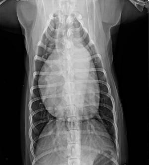
How do all these defects interact in one single heart? The mitral valve dysplasia is mild – there is not much regurgitation and not much stenosis present. So, the left atrial enlargement is certainly caused by the PDA.
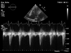
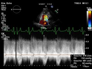
The aortic valve dysplasia creates a stenosis. Still, a large PDA very likely contributes to that stenosis by increasing the diastolic volume of the left ventricle (which has to leave the ventricle through the aortic valve in systole, creating a so called pseudo-stenosis – a stenosis caused by increased volume, not by a reduced diameter). The stenosis is responsible for the thickening of the left ventricular myocardium. The pulmonic stenosis causes right ventricular concentric hypertrophy. Contributing pulmonary hypertension is very unlikely due to the high normal pressure gradient across the PDA. The pressure gradient across the PDA is high normal – On possible explanation would be a mildly decreased pressure within the pulmonary artery due to the pulmonic stenosis. There is reason to assume that the pulmonic stenosis is responsible for the fact that Gordon’s heart has not yet decompensated, meaning that pulmonic stenosis saves him from pulmonary edema. Due to the fact that Gordon did not show any clinical signs of cardiac disease, no medication was prescribed. At the time of this case, catheter-based closure of the PDA and balloon dilation of the pulmonic valve was discussed with the owners but no decision had been made yet.


