Contact Us or call 800-838-4268 for more information
Skip to main content

Prostatic carcinoma diagnosed by FNA: Our Case Of The Month October 2014

Patient Information

Age
15 Years
Gender
Male, Neutered
Species
Canine

Keywords

Blood Chemistry

Clinical Signs

Images

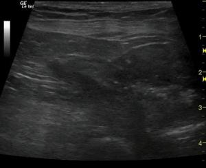
Image 1: The prostate revealed a large mineralizing mass.
Image 2: Extrusion of the mass into the pre-prostatic urethra.
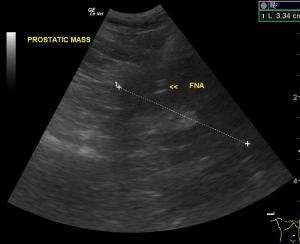
Image 3: FNA of prostatic mass.

So welcome to veterinary life of owner financial constraints of "No" on a UA charge but "Yes" on a sonogram. So this 15 year-old Westie presented for polydipsia and polyuria and suspicion of bladder/urethral stones… well put a probe on it & have a needle in hand & let's just give him bad pathology :( Sneaky prostatic carcinoma at times just pops on the probe when you live by the sonogram as long as you are imaging the prostatic urethra adequately.
Stay tuned because Lindquist and the SonoPath crew may have a palliative solution for this pathology coming in 2015 as we have something up our interventional sleeve. :) Nothing like a probe and a cool set of tools to help out our 4-legged friends with a plumbing problem… and you thought the UGELAB (http://avmajournals.avma.org/doi/abs/10.2460/javma.240.1.51) was cool. :) Stay tuned … but in the meantime check out the 2 minute prostatic carcinoma dx by means of the probe.

History

A 15-year-old MN West Highland White terrier dog was presented for evaluation of polydipsia, polyuria and possible bladder stones noted on radiographs. Abnormalities on CBC and serum biochemistry were thrombocytosis, neutrophilia, and hyperalbuminemia. Urinalysis was not performed owing to financial constraints however an abdominal ultrasound prescription was approved.

Clinical Differential Diagnosis

Polydypsia - bladder pathology (uroliths, neoplasia, chronic cystitis, bacterial cystitis), prostatic neoplasia, renal pathology (early chronic kidney disease, neoplasia, pyelonephritis).

Image Interpretation

The prostate revealed a large mineralizing mass that measured 4.4 x 2.7 cm with extrusion of the mass into the pre-prostatic urethra. Prostatic capsular expansion and distortion is noted.

Sonographic Differential Diagnosis

These images are suggestive of a mineralizing prostatic mass as opposed to luminal urethal calculi. Specifically carcinoma is suspected.

Sampling

U/S guided FNA was performed. Cytology of the prostatic mass revealed carcinoma.

DX

Prostatic carcinoma.

Outcome

Client was considering euthanasia as the patient was not doing well. No further outcome was provided.

Comments

Empirical therapy with Deramaxx and enrofloxacin was recommended until an oncology consultation could be considered.

Videos

Video 1: The prostate revealed a large mineralizing mass.
Video 2: Extrusion of the mass into the pre-prostatic urethra.
Video 3: FNA of prostatic mass.