Contact Us or call 800-838-4268 for more information
Skip to main content

The Shunt Hunt !

Patient Information

Age
8 Months
Species
Canine

Images

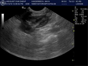
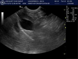
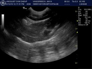
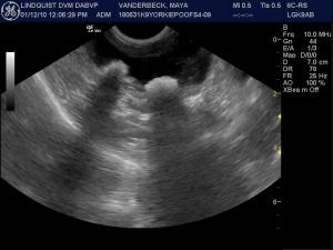
Two separate shadowing cystic calculi are present with minor bladder wall thickening in the near field.
Normal kidney with no significant evidence of mineralization.
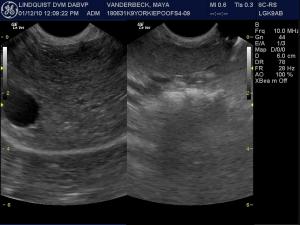
The gall bladder demonstrates normal size and contour. However, the diaphragm takes a sharper angle toward the right of the screen likely indicating subnormal liver size.
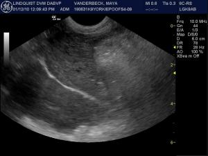
Deep right liver demonstrating mildly coarse parenchyma and normal gastric content to the right.
Right intercostal approach to the liver further demonstrates the coarse and mildly heterogenic parenchyma primarily in the far field.
Right caudal liver demonstrating isoechoic of this portion of the liver when compared to the right kidney.
Transverse view of the liver compared to the normal spleen. Both organs are isoechoic to each other, which is considered normal.
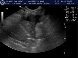
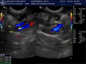
Split portal hilus view demonstrating the portal vein (pv)/vena cava (cvc) ratio on the left and normal portal vein and branches on the right. The 1/1 ratio and normal branching of the portal vein rules out the presence of extrahepatic porto-systemic shunt (i.e. splenocaval, gastrocaval, splenoazygos, gastroazygos shunts).
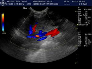
Right intercostal view of the deep vena cava (bottom) and hepatic vein (blue) and artery (red) indicating normal perfusion even thought the liver is subnormal in size. The normal flow and CVC size as well as the normal curvilinear contour of the vasculature rules out the presence of macroscopic intrahepatic shunting.

History

The Shunt Hunt By Sonogram; The nuts and bolts. Does or doesn’t this Yorkie-Poo have a shunt?

Case presented and managed by Dr. Cheryl Welch & staff at Franklin Lakes Animal Hospital, Franklin Lakes, NJ, USA. Sonogram and diagnostic evaluation performed by Eric Lindquist DMV (Italy), DABVP (Canine & Feline Practice).

Sonogram (Liver): Maya

History: An 8-month-old Yorkie Poo was presented at an emergency facility for possible UTI as the owner reported the patient was showing inappropriate urinations in the house, pollakuria, and hematuria. Physical examination was within normal limits. The urine was cloudy and yellow in appearance and on urinalysis was alkaline (pH 8.0) with a normal specific gravity, and leukocyturia (4-10) and a moderate amount of triple phosphate crystals were present. The patient was treated with antibiotics, to which there was no response and he was presented at the RDVM for persistent hematuria a few weeks later. CBC and blood chemistry were both within normal limits. Urine culture was negative for bacterial growth. The only significant finding on survey abdominal radiographs was bladder stones. Abnormalities on repeat urinalysis a week later were high pH (8.5) 4+ proteinuria, 4+, hematuria, and a large amount of triple phosphate crystals. On a bile acids panel the pre-prandial bile acids were normal but the post-prandial bile acids were elevated (post 70 mg/dl) Pending results the patient was placed on the L/D diet.

Clinical Differential Diagnosis

Portocaval shunt Micovascular dysplasia Bladder calculi

Image Interpretation

(Lindquist DMV, DABVP)

Sonographic Differential Diagnosis

Urinary: Cystic calculi and concurrent cystitis. Potential for concurrent urinary tract infection. Liver: Portal vein branch hypoplasia/microvascular dysplasia likely. Non-shunt causes of bile acid elevation also possible such as intestinal dysbiosis or idiopathic transient elevation (See comments below)

Sampling

Surgical liver biopsies were performed which showed arteriolar hyperplasia, portal vein hypoplasia/microvascular dysplasia. Analysis of the bladder calculi (obtained via cystotomy) revealed ammonium biurate calculi.

Outcome

The patient was placed on a medical protocol for microvascular dysplasia, which resulted in resolution of the urinary tract signs.

Comments

(Excerpt from the upcoming textbook Clinical Approach to Sonographic Pathology Lindquist, Frank, and Yanik offered by SonoPath.com in late 2010.) Non-shunt Pathology That Can Elevate Bile Acids Nonhepatic Causes Inflammatory bowel disease/intestinal dysbiosis Spontaneous GB contraction Hypertriglyceridemia Ursodeoxycholic acid treatment Severe disease or resection of the ileum (site of bile acid reabsorption) Cholecystectomy Prolonged anorexia Hyperadrenocorticism Pancreatitis Other nonhepatic pathology Transient elevation in Irish wolfhound puppies, other breeds? Hepatic Causes Diffuse hepatocellular disease Cholestatic disease Primary portal vein hypoplasia/Microvascular dysplasia References: 1. Szatmari V, Rothuizen J, Ultrasonographic identification and characterization of congential portosystemic shunts and portal hypertensive disorders in dogs and cats in Stadards for Clinical and Hostological Diagnosis of canine and Feline Liver Diseases. WSAVA Liver Standardization Group. Saunders, Elsevier Edinburgh, 2006. Pp 15-39. 2. Kirk's Current Veterinary Therapy, Vol XII. Philadelphia: Saunders, 1995:739-740. 3. Willard M, Tvedten H: Small Animal Clinical Diagnosis by Laboratory Methods, 4th ed. St. Louis: Elsevier, 2004241-242. 4. Allen L, Stobie D, Mauldin N, et al: Clinicopathologic features of dogs with hepatic microvascular dysplasia with and without portosystemic shunts: 42 cases (1991-1996). J Am Vet Med Assoc 214, 1999. 5. Gerrizen-Bruning MJ, van den Ingh TS, Rothuizen J: Diagnostic value of fasting plasma ammonia and bile acid concentrations in the identification of portosystemic shunting in dogs. J Vet Intern Med 20:13-19, 2006. 6. Kummeling A, Teske E, Rothuizen J, et al: Coagulation profiles in dogs with congenital portosystemic shunts before and after surgical attenuation. J Vet Intern Med 20:1319-1326, 2006.