Contact Us or call 800-838-4268 for more information
Skip to main content

Splenic Torsion

Patient Information

Age
7 Years
Gender
Female, Intact
Species
Canine

Images

Sampling
Sampling
Sampling
Sampling
Sampling
Sampling

History

Splenic Torsion & The Sonogram.

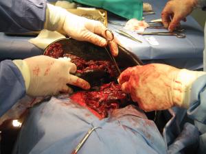
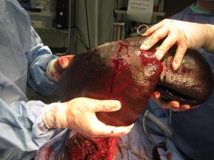
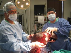
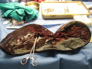
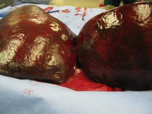
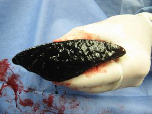
Middle aged Great Dane, vomiting, lethargy, recent onset "ADR"..... put on the probe & ask yourself the clinical sonographer's favorite and most useful question, "What does this?." It's another looks-like-neoplasia-but-isn't pathology imaged & cut by Drs. Cerf & Stancel @ Ridgewood V.H., Ridgewood, NJ, USA & interpretation by Dr. Lindquist of SonoPath.com.

Sonogram (Spleen): Angel Solazo

History(Kohmescer): An intact 7-year-old female Great Dane dog presented for a history of vomiting and lethargic over a couple days. At the previous assessment anemia (32.5%), leukocytosis, and ascites was present. On follow up assessment the anemia had worsened ( 27.5%) and the leukocytosis was similar. ECG revealed VPCs. On cursory abdominal ultrasonography ascites and hyperechoic hepatic nodules were evident. The ascitic fluid was quantified as hemabdomen.

Clinical Differential Diagnosis

(Remo Lobetti PhD, DECVIM): Spleen – neoplasia, torsion, trauma Liver – neoplasia (primary or metastatic), nodular regeneration, hematomas, granulomas Hemorrhagic diathesis Intra-abdominal neoplasia

Sonographic Differential Diagnosis

(Lindquist DMV, DABVP): This is strongly suspicious of a splenic torsion with the potential for underlying, concurrent neoplasia.

Sampling

(Cerf DVM, Stancel DVM, www.ridgewoodvet.com):

Outcome

The patient recovered uneventfully. ECG results were normal on follow-up.

Comments

Special Thanks to Drs. Cerf & Stancel at Ridgewood Veterinary Hospital, Ridgewood NJ, USA for their excellent management of this case. www.ridgewoodvet.com

Videos

The spleen is dramatically enlarged with generalized hypoechoic parenchyma, ill-defined and thickened hyperechoic vascular walls. The perisplenic tissue demonstrates hyperechoic omental changes that appeared to be attached to the spleen. Anechoic ascites is present.
Color flow assessment of the spleen revealed no visible blood flow. Immediate exploratory surgery is recommended owing to suspected splenic torsion and resultant peritonitis.