Contact Us or call 800-838-4268 for more information
Skip to main content

Tetralogy Of Fallot In A 3-Month-Old Female French Bulldog: Our Case Of the Month, March 2022

Patient Information

Age
3 Months
Gender
Female, Intact
Species
Canine

Keywords

Exam Findings

Clinical Signs

Images

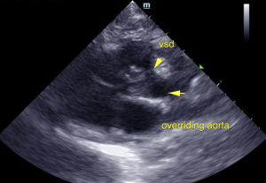
VSD overriding the aorta.
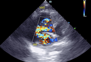
VSD Doppler, MR.
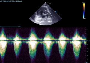
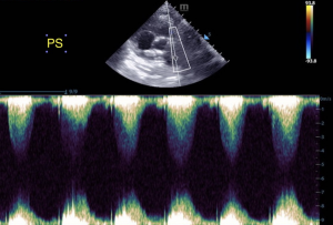
PA Doppler envelopes.
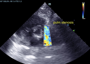
Pulmonic stenosis.
Pulmonic stenosis.

Our own Shari Reffi, CVT, SDEP® certified clinical sonographer for SonoPath Mobile confirms how the SDEP® Echo progression captures even the most complex congenital defects. Tetralogy of fallot: Pulmonic stenosis. Ventricular septal defect. Right ventricular hypertrophy (Right ventricular hypertrophy is a heart disorder characterized by thickening of the walls of the right ventricle.) Overriding aorta (An overriding aorta is a congenital heart defect where the aorta is positioned directly over a ventricular septal defect, instead of over the left ventricle. The result is that the aorta receives some blood from the right ventricle, which reduces the amount of oxygen in the blood.) This complex case was interpreted by Eric Lindquist, DMV, DABVP, Cert. IVUSS.

History

A 3-month-old female French Bulldog puppy presents for a grade 5/6 heart murmur.

Image Interpretation

Concurrent mitral valve insufficiency was noted. The aortic valve was thickened as well and the aorta was overriding. The right atrium was moderately enlarged and measured 1.5:1.0 to the left atrium in four chamber long axis. The tricuspid valve was thickened with insufficiency. Significant right ventricular hypertrophy was noted with stenotic pulmonic valve and right ventricular outflow was severely elevated with a velocity > 6.0 m/sec. Post valvular dilation was noted in the deep pulmonary artery. Membranous ventricular septal defect was noted with thickened, irregular membranous septum.

DX

Tetralogy of fallot. Pulmonic stenosis. Ventricular septal defect. Overriding aorta.

Outcome

Prognosis is poor. The breeding line should be evaluated for similar cardiac defects. Currently, this little heart rock star is under the care of the breeder and living her best life with close monitoring and excellent veterinary care.

Comments

Tetralogy of Fallot includes four defects: Pulmonary Valve Stenosis, Ventricular Septal Defect, Right Ventricular Hypertrophy and Shifting of the Aorta

  • Narrowing of the lung valve (pulmonary valve stenosis). Narrowing of the valve that separates the lower right chamber of the heart (right ventricle) from the main blood vessel leading to the lungs (pulmonary artery) reduces blood flow to the lungs. The narrowing might also affect the muscle beneath the pulmonary valve. Sometimes, the pulmonary valve doesn't form properly (pulmonary atresia).
  • A hole between the bottom heart chambers (ventricular septal defect). A ventricular septal defect is a hole in the wall (septum) that separates the two lower chambers of the heart (left and right ventricles). The hole causes oxygen-poor blood in the right ventricle to mix with oxygen-rich blood in the left ventricle. This causes inefficient blood flow and reduces the supply of oxygen-rich blood to the body. The defect eventually can weaken the heart.
  • Thickening of the right lower heart chamber (right ventricular hypertrophy). When the heart's pumping action is overworked, the muscular wall of the right ventricle becomes thick. Over time this might cause the heart to stiffen, become weak and eventually fail.
  • Shifting of the body's main artery (aorta). Normally the aorta branches off the left ventricle. In tetralogy of Fallot, the aorta is in the wrong position. It's shifted to the right and lies directly above the hole in the heart wall (ventricular septal defect). As a result, the aorta receives a mix of oxygen-rich and oxygen-poor blood from both the right and left ventricles.

Videos

SDEP® position 1.
VSD Doppler.
Pulmonic stenosis Doppler.