Contact Us or call 800-838-4268 for more information
Skip to main content

UGELAB (Laser Ablation) treatment for trigonal transitional cell carninoma of the bladder in a 10 year old FS American Eskimo dog. Our Case Study Of The Month July 2014

Patient Information

Age
10 Years
Gender
Female, Spayed
Species
Canine

Images

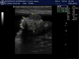
Image 1: Strongly mineralizing dorsocaudal bladder mass occupying the regions of the ureteral papillae. Strongly consistent with transitional cell carcinoma of the bladder.
Image 2: High resolution image of the same bladder mass noting progressive acoustic shadowing owing to the solid density of the bladder mass and strong mineralization.
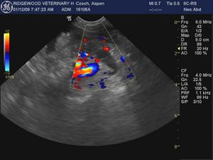
Image 3: Negative color flow uptake on the anechoic dilated left ureter (right in image) as it enters into the bladder trigone. A mineralizing mass is obstructing both ureteral papillae.
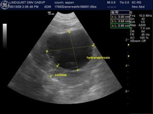
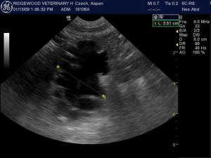
Image 4: Moderate hydronephrosis of the left kidney owing to ureteral obstruction.
Image 5: Severe hydronephrosis of the right kidney owing to distal ureteral obstruction.
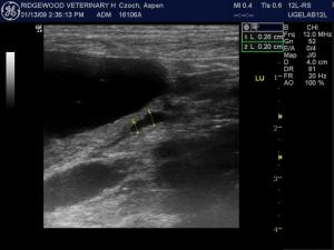
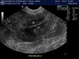
Image 6: Minor residual ureteral dilation after ultrasound-guided laser ablation of the bladder mass that was obstructing the ureteral papillae. Only a minimal amount of residual tumor tags are noted.
Image 7: Post UGELAB procedure: Near-complete resolution of the prior hydronephrosis of the left kidney owing to ureteral obstruction.

Whats wrong with the patient's plumbing? Well, we have a plumber with a really cool set of tools to manage the problem. Obstructive TCC in the urethra? Obstruction of the ureteral papilla? Call the UGELAB plumber at Ridgewood Vet Hospital, Ridgewood NJ. Texas A&M University is doing the UGELAB procedure as well. UGELAB and Stents and SUBs… there are all new plumbing solutions in vet med nowadays but in this case see with Dr. Dean Cerf with Lindquist support did with this challenging urinary plumbing issue by means if Ultrasound Guided Endoscopic Laser Ablation… the UGELAB. You don't have to go to the moon to consult with Dr. Evil & his Alan Parson's Project to obtain access to a "Laser" for this medical procedure. :)

History

A 10-year-old FS American Eskimo dog was presented for stranguria, hematuria, pollajuria and just not herself. She was non responsive to therapy for UTI. Severe azotemia and moderate anemia developed. Urinalysis revealed proteinuria, elevated WBC, blood and transitional cells, and isosthenuria.

Clinical Differential Diagnosis

ARF, CRF, bladder neoplasia, obstructive urolithiasis, resistent UTI, infectious.

Image Interpretation

A 3.5 centimeter mineralizing bladder mass is occupying the trigone and obstructing the ureters at the region of the ureteral papillae. The ureters did not appear invaded by the mass. Mild bladder rotation has occurred owing to mass expansion. Moderate to severe hydronephrosis is present in both kidneys owing to ureteral obstruction. Ultrasound-guided laser ablation was performed for palliative removal of the mass and decompression of the ureters. A minimal amount of ureteral dilation was noted at the end of the procedure with slight residual hydronephrosis on followup. Ureteral function appeared adequate noted by contractility.

Sonographic Differential Diagnosis

Bladder mass with obstructive pattern to both ureters. Non-resectable without ureteral transposition. Strongly mineralizing aspects of the bladder mass suggest transitional cell carcinoma.

Sampling

Endoscopic bx- transitional cell carcinoma

DX

Non-resectable trigonal bladder transitional cell carcinoma with ureteral obstruction.

Outcome

After undergoing the initial UGELAB procedure the patient survived 227 days and then was humanely euthanized.

Comments

Special thanks to Dr. Dean Cerf and staff at Ridgewood Veterinary Hospital, Ridgewood, New Jersey where the ultrasound-guided laser ablation procedure was founded. More information on this procedure may be found in JAVMA Jan1 2012, Cerf D, Lindquist E. (http://avmajournals.avma.org/doi/abs/10.2460/javma.240.1.51?journalCode=javma) or contact Ridgewood Veterinary Hospital directly at 201-447-6000. Texas A&M University, Department of Radiology also performs this procedure

Videos

Video 1: Trigonal bladder mass with ureteral obstruction. Note the dilated ureter as it enters into the trigone. The trigone appears to be ventral in his bladder image however this is owing to the angle of approach of the probe and bladder rotation owing to mass expansion.
Video 2: Separate angle of the same mass further demonstrating ureteral obstruction.
Video 3: Ultrasound-guided laser ablation procedure on the bladder mass. The hyperechoic tissue necrosis line (HTNL) is noted in the bladder mass which indicates ablation of the neoplastic tissue. This tissue will slough over the following days if it is not liberated at the time of the procedure.
Video 4: High resolution of obstructed ureter noted on the right with residual mineralizing bladder mass on the left. This image was taken mid procedure during the UGELAB.
Video 5: End UGELAB procedure that demonstrates only slight ureteral dilation and near complete ablation of the bladder mass. Slight residual tumor is noted to the left of the screen measuring a couple of milimeters. The echogenic tissue represents residual HTNL that will slough in the sequence days.
Video 6: Collapsed right ureter with adequate peristalsis at the end of the procedure.