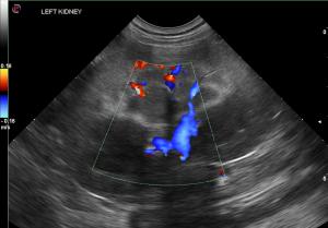
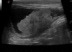
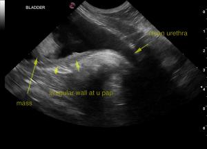
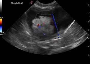
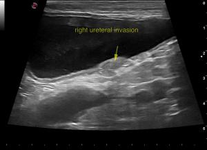
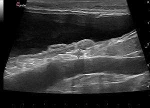
Hematuria, stranguria, pollakuria, and any sort of “uria” needs a probe. Living by “If it's sick it needs a probe” will always provide direction in a case. But when hematuria leads to the sonographic diagnosis of a bladder mass, it's important to define it well sonographically to decide which treatment to pursue, whether surgery or other. With the availability of UGELAB, urinary stents, SUB (subcutaneous ureteral bybass) devices and even artificial neobladders, the clinical sonographer’s job is to define the crucial structures for interventional planning. This is exactly one of those cases where the pelvic urethra, CUJ (cystourethral junction), trigone and ureters have a story to tell when invaded by this carcinoma. Watch Andi Parkinson of Intrapet Imaging. Baltimore, MD, USA show her sonographic footprint while artistically demonstrating this TCC (transitional cell carcinoma) invading the right ureter but still clean in the CUJ and urethra. Probe wielding colleagues I say... this is just how we do it and Andi is setting the bar. :)
DX
Dorsal bladder mass encroaching upon the right ureteral papilla with early obstruction.
Outcome
Ureteral stent placement may be the best option with chemoreduction or surgical reduction of the bladder
mass. Neither kidney revealed pyelectasia at the time of imaging. There was suspicion of developing pyelectasia of the right kidney over time. Invasion of the right ureter appeared to be present in the last 1.0 cm of the ureter.
A guarded to poor long term prognosis was given.

