Contact Us or call 800-838-4268 for more information
Skip to main content

A 1 lb Trichobezoar Foreign Body In A 4-Year-Old MN Feline: Our September Case Of The Month

Patient Information

Age
4 Years
Gender
Male, Neutered
Species
Feline

Keywords

Images

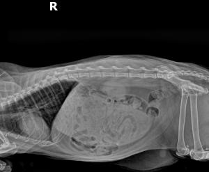
Right lateral abdominal radiograph.
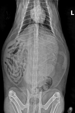
VD abdominal radiograph.
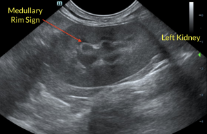
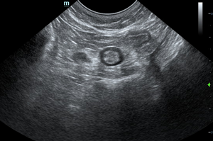
Medullary rim sign in the left kidney- nonspecific.
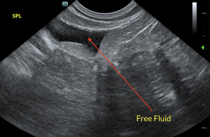
Minor volume free fluid in the area of the spleen.
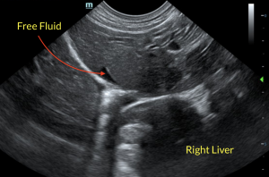
Minor free fluid in the region of the right liver.
Ileocecal valve with prominent colic lymph node located on the left.
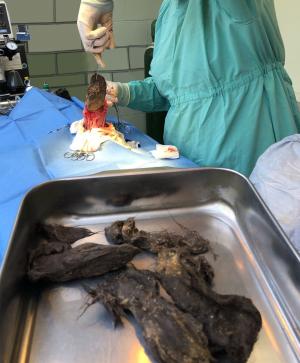
Large trichobezoar being extracted from the stomach during exploratory/gastrotomy.

From fleas to foreign body, how these irriating wingless creatures caused cat barbering (excessively over-grooming to the point where the cat begins to lose hair or creates bald spots in their coat) to the extent that the patient developed a huge trichobezoar in the stomach. Jennavieve Walsh, SDEP® Certified Clinical Sonographer from Animal Sounds NW provided the diagnostic images for this interesting case, detailed interpretation by R. McKenzie Daniel, DVM, DABVP of SonoPath.   Dr. Kelsie Thielen of Faithful Friends Animal Clinic performed the gastrotomy. Post-operative care and management of this case by Dr. Thielen and the compassionate staff at Faithful Friends.

History

A 4-year-old MN DLH cat presented for alopecia; he had fleas at the time and was barbering his hair. A palpable, cranial abdominal mass measuring approximately 4.5 cm x 5 cm was found during exam. The initial thought was that the mass was possibly an enlarged lymph node as it was not painful. At a recheck appointment 2 weeks the later the mass was smaller, felt softer, and the cat had lost 1/2 lb. The patient presented 2 years later due to vomiting, over-grooming of himself and his housemates; a possible splenic mass was a concern. After eliminating evidence of a splenic mass on radiographs, an ultrasound was scheduled for further diagnosis.

Image Interpretation

Left kidney: A hyperechoic corticomedullary band, consistent with a medullary rim sign, was present.The stomach exhibited marked distension containing hyperechoic to progressively shadowing ingesta.

Gastrointestinal: The degree of gastric distension prohibited full evaluation of the gastric walls. The visualized gastric walls were unremarkable. The small intestine presented intact wall layering with 1:3 muscularis/mucosa ratio. The lumen of the small intestine was empty with no signs of ileus, obstruction or foreign material. The duodenum and jejunum wall measured 0.25 cm in width. The ileocolic wall measured 0.38 cm in width.

Free abdomen: Minor volume anechoic free fluid present around the liver and in the lateral abdomen.

DX

Severe gastric distention with retained hyperechoic to progressively shadowing ingesta.

Outcome

The sonographic findings indicated a potential functional gastric motility disorder or gastroparesis. The possibility of non-visualized mechanical obstruction to ingesta outflow could be excluded yet was not obvious given lack of small intestinal mural pathology. No overt evidence of masses or neoplastic criteria was noted. Hospitalization with 24 hour IVF and GI support with documented fast and monitoring for evidence of gastric emptying was recommended. If persistent retained ingesta, gastric emptying via laparotomy with gastrointestinal biopsies may be indicated. The patient underwent an exploratory surgery during which a 1 pound trichobezoar was removed from the stomach via gastrotomy. During follow up exam, the patient was reported to be doing very well.

Videos

Pylorus with foreign material.
Hyperechoic to progressively shadowing ingesta.