Contact Us or call 800-838-4268 for more information
Skip to main content

Abdominal Abscess/Infarcted Lipoma in a 10-year-old MN Beagle: Our Case Of the Month January 2020

Patient Information

Age
10 Years
Gender
Male, Neutered
Species
Canine

Keywords

Clinical Signs

Images

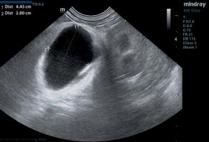
Omental abscess/infarcted lipoma visualized on ultrasound.
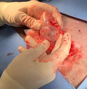
Omental abscess/infarcted lipoma externalized during exploratory surgery prior to removal.
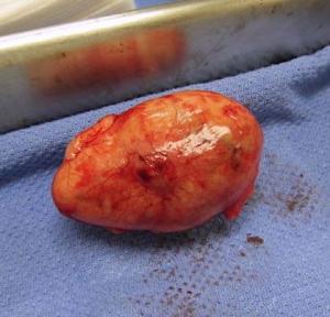
Mass removed.
Patient doing well at suture removal. :)

There are those moments when a clinical sonographer performs a sonogram on that “Ain’t Doin’ Right-ADR” patient that's "just a little off" and we can’t explain why with our traditional outward-in clinical approach. Fortunately for us, we clinical sonographers get to approach a case from the inside-out by means of the probe. As we often say in say in these cases, “Well that isn’t supposed to be there” and off to surgery we go. In this Case Of the Month to kick off 2020 we have the not so uncommon omental abscess/infarcted lipoma that Dr. Murphie ran into with his SDEP™ abdominal protocol. The SDEP™ protocol is made precisely for the ADR patient to image every nook and cranny of the abdomen that also includes pathology not attached to any organ with our “bears in the forest” approach in SDEP™ steps 15-18. Take a look of what is not supposed to be present in this 10-year-old ADR Beagle.

Many thank to Dr. Robb Murphie of VCA Anderson's Corner Animal Hospital for his excellent imaging and management of this interesting case.

History

The patient presented for pre-anesthetic work up for a TPLO surgery and it was noted that the patient's abdomen appeared larger than normal. Radiographs of the abdomen were inconclusive and an ultrasound was performed.

Image Interpretation

Hypoechoic caudal abdominal abscess or infarcted lipoma. Slight fluid accumulation was noted around the abscess.

Sampling

Tru-cut surgical biopsies of the mass were taken during exploratory surgery.

DX

Necrotic adipose tissue with granulomatous inflammation and fibrous encapsulation

Outcome

The caudal abdomen revealed a 4.45 x 2.8 cm hypoechoic abscess or infarcted lipoma with hyperechoic granulation bed and inflammation with slight fluid accumulation noted around the abscess. Surgical intervention was recommended. The patient underwent an exploratory surgery and the mass was remove. The mass was determined to be necrotic adipose tissue with granulomatous inflammation and fibrous encapsulation, most compatible with an intra-abdominal lipoma or strangulated omental fat, which has become necrotic and secondarily inflamed. The patient was found to be doing great and fully recovered at suture removal appointment.

Videos

Microconvex probe imaging of omental abscess/infarcted lipoma
Linear probe imaging of omental abscess/infarcted lipoma