Contact Us or call 800-838-4268 for more information
Skip to main content

Post-Hepatic Obstruction With Chronic Cholangitis In A 12-Year-Old FS Poodle Mix: Our Case Of the Month 2019

Patient Information

Age
12 Years
Gender
Female, Spayed
Species
Canine

Images

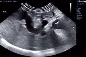
Gallbladder with polyp formation near the neck; note increased portal markings and coarse liver architecture
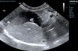
arrow indicating a gallbladder calculus; note hyperechoic outline of the calculus
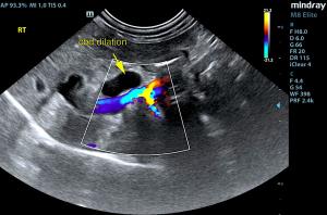
Dilated cystic duct and dilated tortuous common bile duct. Pylorus in near field. The next step is to add color Doppler.
Color Doppler easily distinguishes the portal vein from the common bile duct (no flow) which is normally located between the pylorus and the portal vein in a modified SDEP position 13 view.
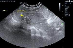
Shadowing calculi in the common bile duct about 2 cm prior to the duodenal papilla. The likely chronic regional inflammation in this area presents a concern for the health of the post-calculus CBD.

Pain, icterus, elevated total bilirubin and liver values - a bell goes off in every sonographer's head over this combo. Effectively imaging the portal hilus region- including the gallbladder, cystic duct, common bile duct, duodenal papilla, pancreas and pancreatic region- is essential and is exactly how Heidi Putnam, SDEP™ Certified Clinical Sonographer with Animal Sounds Northwest, handled the imaging of this case

The patient is under the diligent care of Dr. Bugarovich of VCA Westmoreland Animal Hospital. Patient is fairly stable on medical therapy post scan; surgery is still being considered an option if patient starts to decline.

Want to learn how to image the critical portal hilus as beautifully and throughly as this, so you can nail down a diagnosis? This view is SDEP Position 13, standard in the SDEP protocol and taught in our labs.  For a list of our remaining 2019 labs, click here. Our 2020 lecture/lab dates will be available soon. 

History

The patient was presented for painful abdomen, pyrexia, vomiting, and anorexia. After 12 hours on IVF and supportive care the patient's pyrexia resolved and there was no further vomiting. A painful abdomen persisted and mild icterus was evident in both the sclera and mucous membranes. The patient was treated with Cerenia, Unasyn, Metronidazole, Gabapentin, and Famotidine. Blood chemistry found AST 70, ALT 553, ALK. Phos. 8162, GGT 46, T. Bili. 7.2, K 3.4, Chol. 720, Trig. 609, amylase 2038, PSL 697. Urine specific gravity 1.011, proteinuria 2+, bilirubinuria 3+.

Image Interpretation

The liver revealed coarse architecture and increased portal markings. Gallbladder polyps and calculi were noted. The common bile duct was dilated to 0.6 cm. A shadowing structure was noted at the termination of the common bile duct with tissue thickening or mucoduct personation in the last 2.0 cm of the common bile duct prior to the duodenal papilla. The echotexture of the sludge and shadowing material in the common bile duct would be most consistent with calculus and concurrent thickening of the common bile duct with inflammation. Expansive masses were not present. Acoustic shadowing was noted. The calculus measured approximately 0.4 cm. Grouping of calculi measured approximately 1.0 cm.

DX

Common bile duct calculus. Post hepatic obstruction with chronic cholangitis. Mild potential for common bile duct neoplasia with post hepatic obstruction and hepatic remodeling.

Outcome

Surgical intervention is recommended in this patient with expectations towards bile duct redirection and removal of the calculus. However, the last 2.0 cm of the common bile duct does not look healthy owing to chronic inflammation and likely stricturing. There was no overt evidence of neoplasia; however, there is a minor potential for distal common bile duct neoplasia, yet not suspected.

Videos

It is important to image from the gallbladder, cystic duct, common bile duct to the duodenal papilla to fully examine for the source of the obstructive pattern, as seen here.
Even closer examination of the shadowing calculus and the last 2 cm of the common bile duct leading into the duodenal papilla. This is an important view for surgical planning as the viability/neoplastic potential of this portion of the CBD is in question.